Histopathological Studies of African Yam Bean Seeds and Seedling Infection by Aspergillus niger
-
Chiukpai, Nene Nkata
Department of Plant Science and Biotechnology, Faculty of Science, University of Port Harcourt, Choba, P.M.B. 5323, Port Harcourt, Nigeria
Nwachukwu, Eunice OluchiDepartment of Plant Science and Biotechnology, Faculty of Science, University of Port Harcourt, Choba, P.M.B. 5323, Port Harcourt, Nigeria
Ikechi-Nwogu, Chinyerum Glori

Department of Plant Science and Biotechnology, Faculty of Science, University of Port Harcourt, Choba, P.M.B. 5323, Port Harcourt, Nigeria
| Received 30 Oct, 2024 |
Accepted 01 Jan, 2025 |
Published 13 Jan, 2025 |
Background and Objective: Aspergillus niger is a common fungal pathogen that infects African yam bean seeds and seedlings, leading to significant agricultural losses. Histopathological studies of African yam bean seeds and seedling infection by Aspergillus niger were conducted using light microscopy. The detection and isolation of seed-borne fungi were done using potato dextrose Agar. Materials and Methods: Eighty samples, each of the Light brown TSs 152 accessions, Reddish brown TSs 138, and smooth brown variety of the African yam bean seed were collected from Orie Ugba Market in Umuahia around July, 2021. The Standard Blotter Method was used to inoculate fungal organisms, which were then isolated and investigated for their pathogenicity using a foliar drop method in a greenhouse. Koch’s postulates were confirmed through reinfection of AYB seedlings with spore suspension, dose dependent effects, and disease development for 10 days. Anatomical transverse sectioning was performed on African yam bean seeds and seedlings to determine fungus position and distribution, using rotating microtomes and histological procedures. Means were compared using the Least Significant Difference (LSD) test at a 0.05 significance level. Results: Aspergillus niger, a pathogen, was found in African yam bean seeds, causing necrotic leaf spots, blight, and defoliation. Pathogenicity tests confirmed the infection, and anatomical sections showed a dense mycelium mass in the seed coat, embryo, endosperm, and aleurone layer. The mycelium invaded epidermal cells, progressed through parenchyma tissues, and caused complete degradation within phloem and xylem vessels. The study found mycelium colonization on the epidermis and xylem vessels, leading to vascular tissue disintegration and blockage. Conclusion: The study reveals that Aspergillus niger significantly damages African yam bean seeds by colonizing and degrading vascular tissues. Its pathogenic effects include necrotic leaf spots, blight, and defoliation.
| Copyright © 2025 Nkata et al. This is an open-access article distributed under the Creative Commons Attribution License, which permits unrestricted use, distribution, and reproduction in any medium, provided the original work is properly cited. |
INTRODUCTION
African yam bean seed (AYB), botanically known as Sphenostylis stenocarpa (Hochst ex. A. Rich), is a member of the Fabaceae family. Of the seven key species of this plant, S. stenocarpa is the most valued1. Among the dicotyledons, it is the second largest and one of the most significant economically. The African yam bean (AYB) is a nutritional powerhouse that is particularly high in minerals, carbs, and protein2,3. Additionally, this underutilized crop has significant concentrations of advantageous bioactive substances including flavonoids and polyphenols, which improve general health and well-being4.
African yam beans are a leguminous plant that is widely produced in Western, Eastern, and Central Africa but is largely underutilised5. Due to their nutrition-rich seeds and tubers, the African yam bean (AYB) is recognized as a vital crop for ensuring food security3 in numerous regions where it is cultivated, often utilized to maintain fallow farmlands in preparation for a forthcoming planting season. Also, Adewale and Nnamani2 in their report stated that AYB has been shown to have pharmacological effects, with findings indicating that it can be used to treat conditions such as gout, arthritis, stroke, insomnia, diabetes, measles, stress, and hypertension.
Plant diseases significantly restrict the widespread utilization of AYB6,7. Nevertheless, little is known about the prevalence of the illness and the infectious organisms that attack AYB. The present study aimed at investigating the seed-borne fungi associated with African yam bean seeds and the effect of A. niger infection within the epidermal walls and parenchyma tissue of the plant structure using histopathological techniques.
MATERIALS AND METHODS
Study area: This research work was conducted in the Department of Plant Science and Biotechnology Mycology/Pathology Laboratory and at the Centre for Ecological Studies (Green House) both in the University of Port Harcourt, Rivers State in September, 2021.
Sample collection: Eighty samples each of the light brown TSs 152 accessions, reddish brown TSs 138, and smooth brown variety of the African yam bean seed were collected from Orie Ugba Market in Umuahia around July, 2021.
Isolation of fungi from AYB seeds: To promote fungal growth, organisms derived from the Standard Blotter Method which is approved by the International Seed Health Testing Association8 were inoculated, which involves taking tiny fungal samples and transferring them onto different media. The organisms were then incubated for five to seven days at 25°C. By the conclusion of the seventh day (7), each of the visible mushrooms had been isolated (various species separated) into a newly prepared medium to create a pure culture, or culture that contained only one species. They were then investigated which involved looking at colonies under a microscope and describing their features.
Afterward, planting was done in the greenhouse in preparation for the pathogenicity test. The foliar drop method of the pathogenicity test was used.
Pathogenicity test using Foliar drop method: By Koch’s postulates, there was a reinfection to confirm that the isolated strain was pathogenic. A modified method by Knight and Sutherland9 was adopted where the test isolate spore suspension was used to infect two weeks old AYB seedlings. The spore suspension was diluted with sterile distilled water to observe the dose dependent effects. The seedlings were inoculated using the drop method with the suspension and covered with polythene bags for 24 hrs to maintain high humidity. The inoculated seedlings were then observed for 10 days for disease development and symptoms.
Histopathological studies: Both naturally infected and artificially inoculated African yam bean seeds and seedlings were subjected to anatomical transverse sectioning. To ascertain the position and distribution of the fungus inside the intercellular and intracellular spaces of the AYB seed coat, embryo and endosperm layer, and parenchyma cells, anatomical sectioning was performed using a rotating microtome. To perform histological procedures such as sample fixation, dehydration and embedding, sectioning, staining, and microscopy, Knight and Sutherland9 methods were employed.
Statistical analysis: The observed data was statistically analyzed as one-way ANOVA and was carried out to test the significant difference between the treatments. The technique of analysis of variance of the randomized complete block design was obtained from this study10. The means were compared using the Least Significant Difference (LSD) test at a significance level of 0.05.
RESULTS AND DISCUSSION
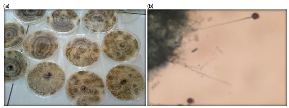
Fungi isolation: The outcome of the fungal isolation was displayed in Fig. 1a and the micrograph in Fig. 1b. From the outcomes of the fungal isolation displayed in Fig. 1a, an unknown fungal species was isolated and discovered to be connected with AYB seeds. Macroscopically, the development of the isolate was white originally but after a few days, it transformed into dark black spores that were visible to ordinary eyes. Based on the photomicrograph, the isolate was known to be an Aspergillus niger. It grows very fast on the PDA media and was dark colored when observed with ordinary eyes and under the microscope, Aspergillus niger had abundantly branched septate, hyaline, and mature hyphae with plenty of conidiophores. The conidiophores remained long and erect each terminating in a rounded head called vesicle in Fig. 1b.
Pathogenicity test: Aspergillus niger was able to infect most of the African yam bean seedlings at different degrees of infectivity four days after inoculation as shown in Fig. 2a and b.
Symptoms showed necrotic leaf spots visible on the leaf surfaces of the infected germinated seedlings. Disease symptoms gradually spread from leaf margins to the midveins, similar to symptoms observed in the African yam bean fields. In a later stage, blight extended to the center of the leaves, and plants became defoliated. Extended necrosis surrounded by yellowing was usually observed on diseased leaves. Pathogenicity tests confirmed that the tested isolate was infectious and no symptoms developed on control plants.
Different degrees of infection on seedlings four days after inoculation.
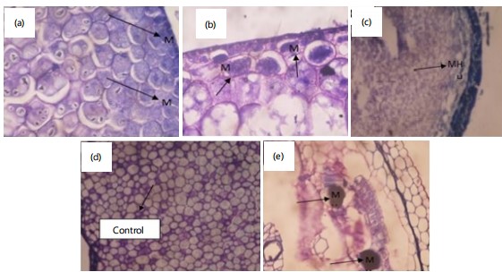
Anatomy of infected AYB seed: Anatomical sectioning of Aspergillus niger-infected African yam bean seeds, both naturally and artificially, revealed that the pathogen’s mycelia mass’s dispersion and colonization throughout the seed coat, embryo, endosperm, and aleurone layer, forming a trichome-like morphology.
|

|
|
A dense mycelium mass of the A. niger pathogen was found in the seed coat, endosperm, and embryonic area of sections of AYB seeds that were naturally and experimentally infected (Fig. 3a-b). Aspergillus niger was widely dispersed throughout the seed coat, endosperm, pericarp, and embryonic portion of the intercellular and intracellular region. showing complete deterioration, cavity, and mycelia mat on the endosperm and the aleurone layer of the cell by mycelium evasion (Fig. 3c and e). The control or healthy seed, showed no signs of fungal dispersion (Fig. 3d).
These results are comparable to the previous research by Gulhane et al.11 and Firdous et al.12, the previous study found that fungi colonize the seed coat, pericarp, and endosperm and embryo region.
|
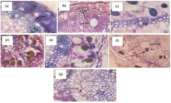
Anatomical sections of the leaves artificially inoculated by A. niger (Fig. 4) revealed that mycelium intensively and extensively invaded intercellular spaces of the epidermal cells and progressed through the parenchyma tissues. (Fig. 4a-g) which showed dense invasion and complete degradation of mycelium within the phloem and xylem vessel. A previous study showed that A. niger infested the host plant African yam bean seedling through the stomata and trichomes13. In the present study, it was found that 4 days after inoculation mycelium was distributed densely in the substomatal spaces of the spongy mesophyll (dark blue mycelium) which is indicated by an arrow. As mycelium spread progressively, the substomatal and intercellular spaces were filled with the pathogen within 4 to 10 days. Figure 4e and f showed that mycelium mat multiplied and colonized in the intercellular spaces. There was no mycelia invasion in the control (Fig. 4g).
The transverse section of AYB infected by Aspergillus niger showed colonization of mycelium on the epidermis and xylem vessels. The dark blue stained mycelium was present in both primary and secondary xylem vessels (Fig. 4). Cambium was also colonized with A. niger mycelium pathogen. The walls of the cambium and phloem were densely colonized with the mycelium mat (Fig. 4e).
Ten days after inoculation, the mycelium invaded and spread to the cortical cells, the petiole, the stem, and the vascular system.
There was distortion and degradation of cortical parenchyma cells (Fig. 4c and d) causing disintegration of the vascular tissue, cortex, and blockage of the xylem and phloem vessels. These results are very much similar to the findings of Nwachukwu and Umechuruba14, who reported the shrinkage of the vascular bundle due to the infection of A. alternata on mango bark 25 days after inoculation.

|
Figure 5 illustrates the transverse sections of the naturally and artificially infected stem tissues of the AYB plant, showing the effects of fungal invasion. Figure 5a depicts the invasion of the xylem vessel by Aspergillus niger mycelium. In Fig. 5b, the xylem vessel is shown to be invaded by fungal mycelium, with noticeable destruction of the cortex. Figure 5c highlights the destruction of the parenchyma cells. Figure 5d presents a healthy transverse section of the stem, showing normal xylem, phloem, and parenchyma cells. Figure 5e shows the mycelium colonization of the xylem vessel and distortion of the cortex 10 days post-inoculation.
The results of this study elucidate the existence of Aspergillus niger in African yam bean (AYB) seeds. Comparable results have been derived from mycological studies concerning AYB seeds found in Nigerian commercial markets. The investigation discerned the presence of various fungi species such as; Aspergillus niger, Aspergillus flavus, Fusarium moniliforme, and Botryodiplodia theobroma as the predominant fungal species found in AYB seeds, thereby corroborating the findings related to the fungi isolated from this present work15.
The findings of the present study are also congruent with earlier research by Khanzada et al.16 that seventeen fungi species belonging to thirteen genera were found to be associated with the African yam bean flower bud and pod and the fungi include: Fusarium verticilloides, Mucor spp., Fusarium oxysporum, Rhizopus spp., Botryodiplodia spp., Penicillium spp., Aspergillus niger, Macrophomina spp., Curvularia spp., Curvularia lunata, Penicillium oxalicum, Pestalotia spp., Cercospora spp., Pythium spp., Colletotrichum spp. and Phomopsis spp.
Fungal pathogens constitute significant impediments to agricultural yield, particularly in Nigeria, where strategies for management are frequently unavailable, economically unfeasible, or not easily attainable by cultivators. There is therefore need for effective surveillance of plant diseases so as to identify possible problems before they generate chief crop losses, while permitting sufficient temporal allocation for the formulation, evaluation, and execution of control strategies.
Aspergillus niger plays a crucial role in the decay of organic matter and contributes to nutrient recycling in ecosystems. It has significant biotechnological importance. It is utilized in various industrial processes. It is generally harmless but can pose health risks to immunocompromised individuals. In some cases, it has been associated with respiratory infections and allergies.
Ochratoxins as a Mycotoxin contamination produced by A. niger poses health risks to humans and animals when contaminated seeds are consumed. Some of these health issues are related to cancer and immunocompromised individuals this is in line with the previous report by Nwachukwu and Umechuruba14.
The effects of reduced germination rates and seedling establishment directly affect crop yields and also contaminated seeds may be rejected for commercial purposes leading to economic losses for farmers.
In contrast to the control, which exhibited no symptoms throughout the pathogenicity test, spots quickly developed on the leaf surfaces and necrosis spread across the entire leaf in four to ten days. After reisolating the injected pathogen, it was discovered that its morphology resembled that of the original isolate.
The pathogenicity result by A. niger was proven to cause an externally and internally seed-borne pathogen leading to local and systemic transmission17.
The fungus penetrated the seed coat and colonized the cotyledons, leading to cell wall degradation where fungal enzymes such as celluloses and pectinases, degrade cell walls causing tissues to collapse.
The pathogenicity test confirmed A. niger as a highly virulent pathogen affecting AYB seeds and seedlings. Seeds inoculated with A. niger displayed reduced germination rates, discoloration, and softening of the seed coat, this is consistent with findings of Dabiré et al.17, who reported that A. niger significantly decreased seed germination. Also, the penetration of the fungus caused nutrient depletion that resulted to the decomposition of starch granules and proteins. These results are in accordance with another study, that Fungi growing on seed reduce germination rate, carbohydrate, protein, and total oil contents and influence other biochemical changes in grains18.
Histological sectioning carried out to study the anatomical and cellular effects of fungal infection on African yam bean (AYB) seeds and seedlings showed positive results. When investigating seeds inoculated with Aspergillus niger either naturally or artificially it provides insight into the fungal invasion, tissue damage, and seed defense mechanisms. Thus, histopathological studies revealed that Aspergillus niger infects the seed and colonizes in various seed tissues and damage the embryo and affect seed viability.
The examination of various anatomical transverse sections of the African yam bean seeds and seedlings using a light microscope revealed histopathological differences between the healthy and the diseased African yam bean tissues.
The extensive colonization results in structural disorganization of tissues, including the breakdown of parenchyma cells.
In seedlings, A. niger colonized vascular tissues, impairing water and nutrient transport, resulting in symptoms like wilting, chlorosis, and stunted growth. These symptoms indicate systemic infection, as the fungus spreads beyond localized tissues into the vascular system.
Histological analysis serves as a foundational tool to understand fungal-seed interactions and their implications for seed quality and agricultural productivity18.
The significance of the histopathological sectioning is that in disease mechanism it helps to elucidate how A. niger colonizes and damages AYB seeds and seedlings. In seed defense, it reveals the anatomical and cellular changes in response to fungal invasion. In breeding programs, it can give an insight from the histological studies and guide on the development of resistant AYB cultivars.
CONCLUSION
This study provides the first comprehensive histopathological report on A. niger infection in African Yam Bean seeds and seedlings. The results show that browning of tissues and lesions occurs only at the infection sites, reducing seed germination and seedling emergence. Aspergillus niger mycelium colonizes densely at both inter and intracellular layers, causing low yield in African Yam Bean production and storage. Leaf spots symptoms appear due to infected mesophyll tissues, disintegrating mesophyll cells, and damaging chloroplast membranes.
SIGNIFICANCE STATEMENT
This study underscores the significant impact of Aspergillus niger infection on African yam bean seeds and seedlings, highlighting its potential to disrupt crop health and yield. The findings emphasize the need for effective disease management strategies to safeguard food security and support agricultural development, particularly in regions reliant on this vital crop.
REFERENCES
- Ojuederie, O.B., M.O. Balogun, S.R. Akande, S. Korie and T. Omodele, 2015. Intraspecific variability in agro-morphological traits of African yam bean Sphenostylis stenocarpa (Hochst ex. A. Rich) harms. J. Crop Sci. Biotechnol., 18: 53-62.
- Adewale, B.D. and C.V. Nnamani, 2022. Introduction to food, feed, and health wealth in African yam bean, a locked-in African indigenous tuberous legume. Front. Sustainable Food Syst., 6.
- Gbenga-Fabusiwa, F.J., 2021. African yam beans (Sphenostylis stenocarpa): A review of a novel tropical food plant for human nutrition, health and food security. Afr. J. Food Sci., 15: 33-47.
- George, T.T., A.O. Obilana and S.A. Oyeyinka, 2020. The prospects of African yam bean: Past and future importance. Heliyon, 6.
- Uchegbu, N.N. and N.F. Amulu, 2015. Effect of germination on proximate, available phenol and flavonoid content, and antioxidant activities of African yam bean (Sphenostylis stenocarpa). World Acad. Sci. Eng. Technol. Int. J. Nutr. Food Eng., 9: 106-109.
- El Khoury, W. and K. Makkouk, 2010. Integrated plant disease management in developing countries. J. Plant Pathol., 92: S35-S42.
- de Souza, R.S., J.L.B. Lopes, C.F.R. Geyer, L. da Rosa Silveira João and A.A. Cardozo et al., 2019. Continuous monitoring seed testing equipaments using internet of things. Comput. Electron. Agric., 158: 122-132.
- Oyedele, T.A., I.A. Kehinde, A.S. Oyelakin, T.O.S. Popoola, H.Y. Atanda and L.A.J. Mur, 2024. Identifying the fungal diseases of African yam bean (Sphenostylis stenocarpa) and their incidence in South-West Nigeria. Physiol. Mol. Plant Pathol., 130.
- Knight, N.L. and M.W. Sutherland, 2011. A rapid differential staining technique for Fusarium pseudograminearum in cereal tissues during crown rot infections. Plant Pathol., 60: 1140-1143.
- Gomez, K.A. and A.A. Gomez, 1984. Statistical Procedures for Agricultural Research. 2nd Edn., John Wiley and Sons Inc., Hoboken, New Jersey, ISBN: 978-0-471-87092-0, Pages: 704.
- Gulhane, A.R., S.V. Khambalkar and G.K. Giri, 2018. Histopathological studies of seed infected with seed borne fungi. Int. J. Curr. Microbiol. Appl. Sci., 7: 4108-4112.
- Firdous, S.S., R. Asghar and G. Murtaza, 2014. Histopathology of leaf spot of sesame (Sesamum indicum L.) caused by Pseudomonas syringae pv. sesami. J. Anim. Plant Sci., 24: 814-819.
- Jason, L.A., M.T. Ropacki, N.B. Santoro, J.A. Richman and W. Heatherly et al., 1997. A screening instrument for chronic fatigue syndrome: Reliability and validity. J. Chronic Fatigue Syndr., 3: 39-59.
- Nwachukwu, E.O. and C.I. Umechuruba, 2001. Antifungal activities of some leaf extracts on seed-borne fungi of African yam bean seeds, seed germination and seedling emergence. J. Appl. Sci. Environ. Manage., 5: 29-32.
- Martinelli, J.A., C.A.C. Bocchese, W. Xie, K. O'Donnell and H.C. Kistler, 2004. Soybean pod blight and root rot caused by lineages of the Fusarium graminearum and the production of mycotoxins. Fitopatologia Bras., 29: 492-498.
- Khanzada, M.A., A.M. Lodhi and S. Shahzad, 2004. Pathogenicity of Lasiodiplodia theobromae and Fusarium solani on mango. Pak. J. Bot., 36: 181-189.
- Dabire, T.G., S. Bonzi, I. Somda and A. Legreve, 2016. Identification of seed-borne fungi of onion (Allium cepa L.) in Burkina Faso. Int. J. Innovation Sci. Res., 25: 562-571.
- Bhattacharya, K. and S. Raha, 2002. Deteriorative changes of maize, groundnut and soybean seeds by fungi in storage. Mycopathologia, 155: 135-141
How to Cite this paper?
APA-7 Style
Nkata,
C.N., Oluchi,
N.E., Glori,
I.C. (2025). Histopathological Studies of African Yam Bean Seeds and Seedling Infection by Aspergillus niger. Research Journal of Botany, 20(1), 38-46. https://doi.org/10.3923/rjb.2025.38.46
ACS Style
Nkata,
C.N.; Oluchi,
N.E.; Glori,
I.C. Histopathological Studies of African Yam Bean Seeds and Seedling Infection by Aspergillus niger. Res. J. Bot 2025, 20, 38-46. https://doi.org/10.3923/rjb.2025.38.46
AMA Style
Nkata
CN, Oluchi
NE, Glori
IC. Histopathological Studies of African Yam Bean Seeds and Seedling Infection by Aspergillus niger. Research Journal of Botany. 2025; 20(1): 38-46. https://doi.org/10.3923/rjb.2025.38.46
Chicago/Turabian Style
Nkata, Chiukpai,, Nene, Nwachukwu, Eunice Oluchi, and Ikechi-Nwogu, Chinyerum Glori.
2025. "Histopathological Studies of African Yam Bean Seeds and Seedling Infection by Aspergillus niger" Research Journal of Botany 20, no. 1: 38-46. https://doi.org/10.3923/rjb.2025.38.46

This work is licensed under a Creative Commons Attribution 4.0 International License.